Sporotrichosis
Sporotrichosis is a chronic infection of the fungus Sporothrix
schenckii (S. schenckii) that usually affects the
skin (although it has other, rare forms that affect the lungs,
joints and bones).
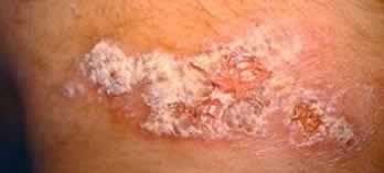
Sporotrichosis lesion.
Sporothrix schenckii Fungus
This fungus is found worldwide and is naturally present in soil,
hay, rose thorn, sphagnum moss, decaying vegetation, and other
plant materials, and infects the skin through small cuts and punctures.
Because of this, this disease usually affects people who work
with soil and plants, such as farmers, horticulturists, and gardeners.
Because of this occupational exposure, sporotrichosis is more
prevalent in adult males.
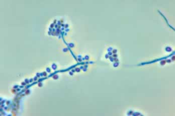
Sporothrix schenckii (S. schenckii) fungus.
In the rarer, pulmonary form of sporotrichosis, the infection
took place when S. schenckii is inhaled.
A rare form of transmission of this fungus is through cats and
armadillos. Sporotrichosis is not communicable from person to
person.
Forms of Sporotrichosis
Sporotrichosis has 3 forms:
- Cutaneous lymphatic sporotrichosis
Red, nodular lesions of the skin, along with secondary lesions
of the lymphatic vessels.
- Pulmonary sporotrichosis
This is a rare form of sporotrichosis that causes nodules in
the lungs.
- Disseminated sporotrichosis
A rare form of sporotrichosis that causes arthritis, osteomyelitis
or inflammation of the bone (osteoarticular sporotrichosis),
and also infects the central nervous system and brain (called
sporotrichosis meningitis). This usually occurs only in people
with weakened immune system.
In disseminated sporotrichosis, the infection spread from the
primary location (skin or lung) to other areas of the body.
This form of sporotrichosis infection is a dangerous and even
life-threatening medical condition.
Signs and Symptoms
Symptoms of cutaneous or common sporotrichosis:
- Small, painless bump or lesion
The S. schenckii infection progresses slowly, with incubation
time anywhere between 1 week and 3 months – the first
symptom is usually a small bump in the skin the looks like an
insect bite. This bump is usually found in the finger, hand,
or arm where the fungus enters through the skin.
- Red to purple in color
The bump can range in color from pink or red to purple. Although
it is usually painless, it can be slightly tender.
- Ulceration
The lesion will then grow larger, discolor, and may even develop
into an ulcer or open up to look like boil and drain clear fluid.
Left untreated, this ulcer can become chronic and persist for
years.
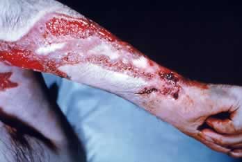 Severe ulceration in sporotrichosis.
- Lesions along the lymp nodes and vessels
Additional bumps may develop and may form along lymph nodes
and vessels.
Left untreated, secondary bacterial infections such as cellulitis
can develop.
Symptoms of pulmonary sporotrichosis include:
- Cough
- Holes and nodules in the lungs
- Swollen hilar lymph nodes of the lungs
Pleural effusion or pooling of blood or lymph fluids into the
pleural cavity. (A pleura is the thin membrane that envelops
the lungs and fold back as a lining for the chest cavity).
- Fibrosis or formation of abnormal fibrous tissue
- Formation of fungus ball
In many patients, sarcoidosis (lesions in the liver, lungs, skin,
and lymph nodes) and tuberculosis can also develop.
Symptoms of disseminated sporotrichosis include:
- Weight loss
- Loss of appetite or anorexia
- Bony lesions
This form of the disease can lead to or be accompanied by arthritis
and osteomyelitis or infection of the bones.
Diagnosis
Due to its subtle and slow-growing nature, diagnosis of S. schenckii
infection can be difficult. A confirming diagnosis is done through
culturing S. schenckii fungus in the pus, sputum (saliva, phlegm,
mucus or spit), synovial tissue biopsy, and arthrocentesis or
bone drainage.
Antibody against S. schenckii can also be used – however,
due to variability in sensitivity and specificity, it cannot be
used as the sole basis for diagnosis. Higher antibody titer in
the cerebrospinal fluid as compared to blood or serum antibody
against S. schenckii suggests the presence of sporotrichosis meningitis.
In case of pulmonary sporotrichosis, due to similarities in symptoms,
other diseases such as tuberculosis and sarcoidosis must be ruled
out. Similarly, in cases of disseminated sporotrichosis, osteomyelitis
due to bacterial infection and bone cancer must be ruled out.
Treatment
Treatment of sporotrichosis on the skin includes:
- Saturated potassium iodide
Skin lesions are treated with saturated potassium iodide solution,
usually given for 3 to 6 months and usually continued for 1
to 2 months after lesions have completely healed.
- Itraconazole
An antifungal medication, itraconazole inhibits fungal cell
growth. In cases where the patients cannot tolerate this medicine,
flucanozole can be used.
- Excision and drainage
In some cases, lesions must be excised and drained.
- Heat
Warming up the affected area may bring temporary relief to the
pain
Treatment of disseminated sporotrichosis includes:
- Itraconazole
Patients may need to take this antifungal medication for several
months, even up to one year.
- Amphotericin B
This is an intravenous drug with serious potential side effects,
including fever, chills, nausea, and vomiting.
- Surgery
Surgery may be required to remove the infected bone.
Treatment of pulmonary sporotrichosis includes:
- Itraconazole
- Amphotericin B
- Surgery
Surgery may be required in cases of cavitary pulmonary lesions
(lesions in the lungs that cause cavities or holes)
Treatment of sporotrichosis meningitis include:
- Amphotericin and flucytosine
- Itraconazole
References:
